

Inside a Brain, Cell by Cell: What Alzheimer's Research Looks Like at Single-Cell Resolution
The brain is made up of billions of cells, but they do not all behave in the same way.
That becomes especially important when researchers study complex diseases such as Alzheimer’s. A neuron may respond differently from a microglial cell or an astrocyte, even when they are exposed to the same disease environment.
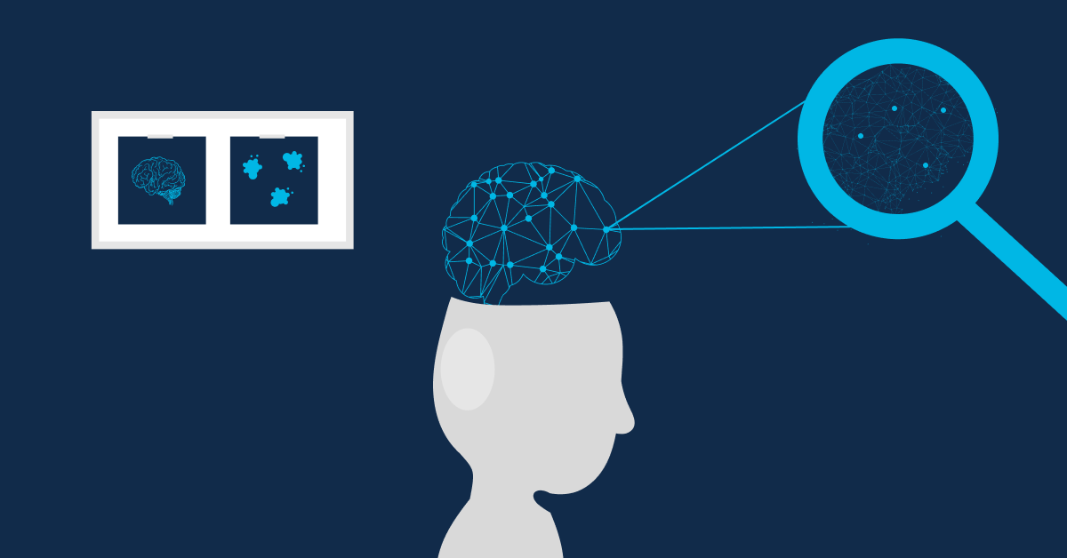
So instead of asking only “Which genes change in an Alzheimer’s brain?”, researchers are increasingly asking:
“Which genes change, in which cells, and at what stage of disease?”
This is where single-cell and single-nucleus transcriptomics can provide a much closer look.
Why Look at the Brain Cell by Cell?
Traditional bulk RNA sequencing measures gene expression across a mixture of cells. This provides a useful overall picture, but cell-specific signals can sometimes become hidden within the average.
Single-cell transcriptomics helps separate these signals by examining individual cells or nuclei.
Researchers can use it to explore:
- Different cell populations within brain tissue
- Gene-expression patterns in specific cell types
- Cellular states associated with disease
- Differences between affected and unaffected samples
- Biological pathways that may change between cell populations
This difference is also why choosing between bulk and single-cell approaches depends on the research question. Our guide on Single-Cell vs Bulk Transcriptomics explores this distinction in more detail.
A 2026 Study Looks Across Cell Types and Populations
A recent 2026 study published in Nature provides a strong example of where this field is heading.
Researchers used single-nucleus RNA sequencing together with chromatin-accessibility profiling to study post-mortem brain tissue from 167 individuals across three population groups and three brain regions.
The study identified Alzheimer’s-associated molecular signatures across several cell types, including:
- Microglia
- Astrocytes
- Neurons
- Oligodendrocytes
The researchers also observed shared as well as region-specific molecular patterns.
That matters because Alzheimer’s does not necessarily produce one uniform molecular response throughout the brain.
Different cell populations and different brain regions can tell different parts of the disease story.
Read the research: Cell-type signatures of Alzheimer’s disease shared across population groups
Microglia Tell Another Part of the Story
Another fascinating 2026 Nature Medicine study focused on microglia, the immune cells of the brain.
Researchers combined spatial transcriptomics with single-nucleus RNA sequencing to examine human brain tissue and investigate cellular changes around the transition from amyloid-β-associated pathology toward tau-associated changes.
The study identified different tissue states and changes in microglial programs along this pathological continuum.
This illustrates why looking at individual cellular states can be useful. Rather than treating all microglia as one population, researchers can investigate:
- Different microglial states
- Molecular changes associated with pathology
- Where particular cellular states occur
- How cellular programs change across disease contexts
Read the research: Human microglial transitions at the Aβ–tau inflection point
What Happens to the Data?
Finding these patterns requires much more than sequencing.
Single-cell datasets can contain thousands or even millions of individual cells or nuclei. Researchers therefore need computational workflows to organize and interpret the data.
Common analysis steps include:
- Quality control to identify reliable data
- Normalization to make expression measurements comparable
- Dimensionality reduction to explore complex datasets
- Clustering to identify groups of similar cells
- Cell-type annotation to understand what those groups represent
- Differential expression to identify genes that vary between groups
- Functional enrichment to investigate biological pathways
- Visualization to make cellular patterns easier to interpret
This is where bioinformatics connects sequencing data with biological questions.
GenomeBeans' Single-Cell Transcriptomics workflow brings together key stages of single-cell data analysis, including quality control, dimensionality reduction, clustering and annotation, differential expression, functional enrichment and visualization.
From Millions of Cells to Biological Questions
The value of single-cell transcriptomics is not simply that it produces more data.
It allows researchers to separate biological signals that might otherwise be mixed together.
In Alzheimer’s research, this can mean exploring:
- How neurons differ from glial cells
- Which cellular populations show disease-associated changes?
- How microglial states vary
- Which molecular pathways are altered
- How cellular responses differ between brain regions
And recent research shows that combining single-cell or single-nucleus approaches with spatial profiling can provide an even richer picture of what is happening within tissue.
Seeing Alzheimer's at Cellular Resolution
Alzheimer’s is a complex disease involving many cell types and molecular processes.
Single-cell transcriptomics helps researchers see that complexity at a much finer resolution.
Instead of looking at one averaged signal from an entire tissue sample, researchers can examine individual cellular populations and ask more specific biological questions.
As technologies and computational methods continue to develop, this approach is helping researchers explore cellular heterogeneity, disease-associated gene programs, regional differences, and changing cellular states.
The more closely researchers can see the individual cells, the more detailed the biological story becomes.
Popular Post
-
14 September 2026
When Mitochondria Fail: How Variant Calling Supports Mitochondrial Disease Research
-
07 September 2026
From Bacteria to Biomarkers: How the Gut Microbiome Is Becoming a Clinical Tool

-
04 September 2026
Beyond Gut Bacteria: How Metagenomics Is Decoding the Human Microbiome

-
26 August 2026
Beyond SNVs: Why Structural Variants and CNVs Are the Missing Piece in Many Genetic Diagnoses

-
17 August 2026
Why Biotech Startups Should Outsource Bioinformatics: ROI, Speed & Scalability